Ureterocele
A ureterocele: what is it?
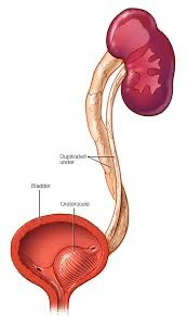
One hereditary disorder that causes the bottom of your ureter to protrude like a balloon within your bladder is called a ureterocele. The tubes that transport urine from your kidney to your bladder are called ureters. Vesicoureteral reflux, or pee backing up to your kidneys, may occur when a ureterocele obstructs the passage of urine. Depending on the nature and location, ureterocles may have a range of sizes and symptoms.
Ureterocele types
A unilateral ureterocele affects just one ureter, whereas a bilateral ureterocele affects both ureters. The more prevalent kind of ureterocele is unilateral.
A ureterocele may also be categorized by medical professionals based on where it is located. Up to 75% of ureterocles are extravesical in nature. Infants and children are most often affected with extravesical ureterocele. It indicates that the bulge reaches the urethra and the bladder neck, which is the bottom of the bladder.
In fewer than 25% of instances, ureterocles may also be intravesical. This kind of protrusion just shows up within the bladder; it doesn’t go outside or to the neck of the bladder.
Causes and Symptoms
What signs of a ureterocele are present?
Except in cases when the disease results in a urinary tract infection (UTI), ureterocles often show no symptoms. A UTI’s symptoms include:
- burning while urinating and/or painful urination.
- pee that smells bad.
- You may have a lump or pain in your abdomen.
- You have blood in your urine.
- urinating more often.
- unable to contain your urination.
- having trouble emptying your bladder completely.
Why does a ureterocele occur?
When the end of the ureter that enters the bladder does not form properly during fetal development, a ureterocele is created. Sometimes the reason for a ureterocele is not apparent until adulthood, but the symptoms are the same.
What issues might arise from a ureterocele?
- Urine may back up into your kidneys when it doesn’t leave your body (urinary retention). Kidney damage may result from this urinary tract infection (UTI).
- Kidney stones are among the additional hazards.
- injury to the bladder.
- recurring urinary tract infections.
Once kidney function is compromised, it cannot be restored because kidney damage is permanent. Therefore, it’s critical to get therapy in order to maintain renal function.
Testing and Diagnosis
How does one diagnose a ureterocele?
Numerous ureteroceles may be identified by medical professionals using a prenatal ultrasound. Usually, this happens around week 20 of pregnancy. Your doctor may suspect a ureterocele if the ultrasound shows swelling in the kidney and ureter. It may be verified by an imaging examination after delivery. Sometimes a diagnosis of ureterocele is made after delivery. However, by the age of two, the majority of kids with a ureterocele are diagnosed.
However, the syndrome is still present in elderly individuals and children who have a urinary tract infection or kidney stones. Any of the following tests might be ordered by your provider:
- an ultrasonography of the kidney.
- a voiding cystourethrogram (VCUG), a test for the bladder.
- a kidney scan to assess kidney health.
- To examine the kidneys and bladder, use magnetic resonance imaging (MRI) and computed tomography (CT) scans.
- a urine test to detect an infection of the urinary tract.
How may a ureterocele be fixed?
The following factors determine how a ureterocele is treated: When medical professionals identify it.
- if your kidneys are impacted.
- if vesicoureteral reflux (VUR) is present.
In order to lower the risk of infection until surgery, your doctor may prescribe antibiotics if a prenatal ultrasound reveals it. The best course of action will be decided by your child’s healthcare practitioner after birth.
Watchful waiting or surgery are further therapy possibilities.
Surgical procedures
Surgery is performed to prevent the ureter from enlarging and becoming blocked. Your physician may use one of the following surgical techniques:
- Transurethral puncture: The ureterocele is punctured during this surgery. A cystoscope is inserted by a surgeon into the bladder and urethra to do this. This process is similar to bursting a balloon. Although it is often performed as an outpatient treatment, which eliminates the need for an overnight hospital stay, general anesthesia is still used, which puts the patient to sleep throughout the process.
- Ureteric Re-implantation: In order to access the bladder during ureteric reimplantation, a surgeon must make an incision in the lower abdomen. The ureter is then properly reimplanted into the bladder after the surgeon removes the ureterocele. In order to stop VUR (urine reflux), they additionally reconstruct the flap valve. Although this surgery is often more complicated, it is effective 90–95% of the time.
- Upper polar Nephrectomy : When the upper portion of your kidney stops functioning because of damage from a ureterocele, you may need an upper pole nephrectomy. People who have a duplex kidney with a ureterocele are more likely to experience this.
A vigilant waiting
A healthcare professional who practices watchful waiting keeps an eye on your condition over time to track its development. If you’re an adult or if there doesn’t seem to be any kidney damage and your ureterocele isn’t generating any symptoms, this could apply to you.