Testicular Cancer
Testicular cancer: what is it?
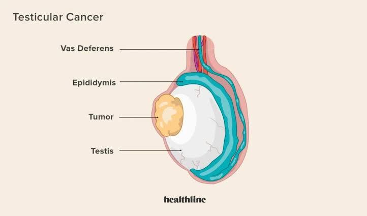
When malignant (cancer) cells grow in the tissues of one or, less often, both testicles, testicular cancer results. Sperm and the hormone testosterone are produced by your testicles, which are two walnut-shaped sex glands. They are located within the scrotum, a skin sac underneath your penis.
Testicle cancer is a dangerous disease, much like any other cancer. Thankfully, testicular cancer is very curable and treatable.
Which forms of testicular cancer exist?
The germ cells in your testicles that aggregate to create a mass or tumor are the cause of around 90% of all testicular cancers. Eventually, germ cells mature into sperm. Germ cells are the source of two forms of testicular cancer.
- Seminoma: A cancer that grows slowly and usually strikes adults in their 40s or 50s.
- Non-Seminoma – Cancer that develops more quickly than seminomas is called a non-seminoma. People in their late teens, 20s, and early 30s are most affected. Non-seminoma tumors come in four different varieties. Each has a name derived from the kind of germ cell that creates the tumor. Teratoma, choriocarcinoma, yolk sac carcinoma, and embryonal carcinoma are examples of non-seminoma cancers.
Both seminoma and non-seminoma cells may be seen in certain testicular cancer tumors.
What is the prevalence of testicular cancer?
Only about 1 in 250 persons with testicles may get testicular cancer. Nonetheless, it is the most prevalent malignancy in men between the ages of 15 and 35.
Causes and Symptoms
What symptoms may one expect from testicular cancer?
- A painless lump in your testicle is the most typical indication of testicular cancer. Other signs and symptoms include:
- scrotal swelling or an unexpected accumulation of fluid.
- an enlargement or lump in one or both testicles.
- a sensation that your scrotum is heavy.
- dull pain in your lower abdomen or groin.
- discomfort or pain in a testicle or scrotum.
- a testicle that is becoming smaller (testicular atrophy).
Testing and Diagnosis
How is the diagnosis of testicular cancer made?
After looking at a lump or other change you discovered in your testicle during a self-examination, your doctor can conclude that you have testicular cancer. Testicular cancer may sometimes be discovered during a standard physical examination.
Testicular cancer is often diagnosed using the following methods and tests:
- A history and physical examination: In order to look for indications of testicular cancer, your doctor will question you about your symptoms and do a thorough examination. They could check your lymph nodes for indications of cancer spread and feel your testicles for lumps.
- Ultrasound: Your doctor will probably request an ultrasound if they find anything unusual during the examination. Using high-energy sound waves, an ultrasound is a harmless medical treatment that produces images of the tissues within your body.
- Inguinal orchiectomy and biopsy: Your doctor will make an incision (cut) in your groin to remove the afflicted testicle if the ultrasound reveals signs of malignancy. A expert will use a microscope to look for cancer cells in testicular tissue.
Additional testing might consist of:
- A serum tumor marker test measures the levels of certain chemicals associated with particular cancer types by analyzing a blood sample. We refer to these compounds as tumor markers. Alpha-fetoprotein (AFP), human chorionic gonadotropin (HCG or beta-HCG), and lactate dehydrogenase (LDH) are tumor markers that are often increased in testicular cancer. Different markers are elevated by different tumor types. Seminomas, for instance, may sometimes increase HCG but not AFP. HCG is not raised by non-seminomas, although AFP is. Increased LDH levels might be a sign of cancer metastasis.
- MRIs, CT scans, and X-rays: X-rays are used in a CT scan to create images of your inside organs. To determine if your cancer has progressed to your abdominal organs, your doctor could undertake a CT scan of your abdomen and pelvic. To determine if cancer has spread to your lungs, they could prescribe a CT scan or a routine X-ray. You could get an MRI if your doctor believes that cancer has progressed to your brain and spinal cord. An MRI creates images of the interior of your body using radio waves and magnets.
Which testicular cancer stages are there?
Cancer staging is another aspect of diagnosis. Important details like tumor size and whether the disease has spread are provided by staging, which can help dictate therapy choices.
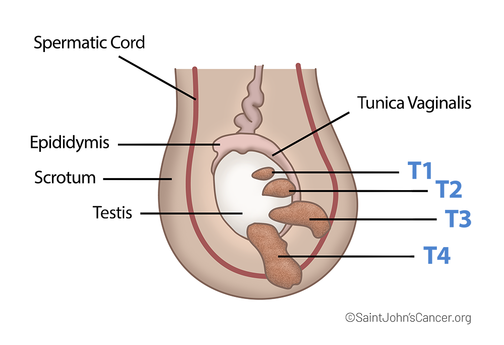
- Stage 0: Although abnormal cells have grown, they are still within the testicles, which are where sperm cells begin to form. Germ cell neoplasia in situ (GCNIS) is another name for stage 0.
- Stage I: The cancer is limited to the testicle, maybe encompassing adjacent lymphatic or blood arteries. There may or may not be increased tumor markers.
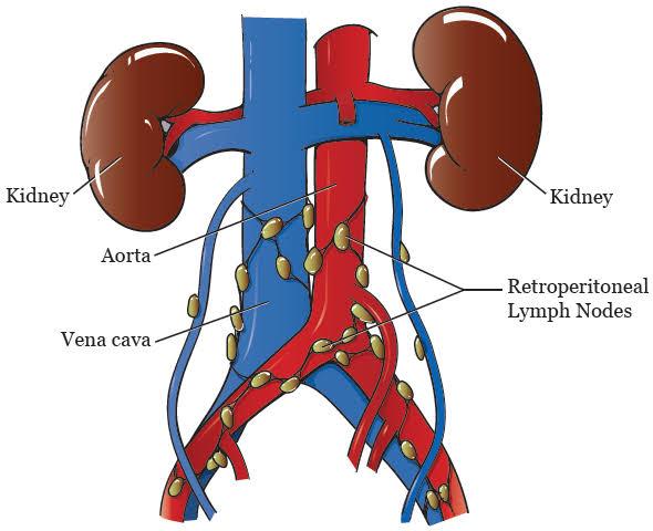
- Stage II: The cancer has only reached the retroperitoneum, or rear of the belly, in terms of lymph nodes. You are in stage III instead of stage II if you have lymph node cancer and moderately or significantly increased tumor markers.
- Stage III: The cancer has progressed to an organ or lymph nodes outside of the abdomen.
Treatment
What therapies are available to treat testicular cancer?
Your health, treatment choices, cancer stage, and tumor kind are some of the variables that affect how you are treated. Compared to non-seminomas, seminomas often develop more slowly and react better to radiation treatment. Chemotherapy works effectively for both types of testicular cancer tumors.
Your doctor will treat testicular cancer as a non-seminoma if it has both seminoma and non-seminoma tumors.
Operation
Regardless of the tumor kind or disease stage, the most frequent therapy for testicular cancer is surgery to remove the malignant testicle. Your healthcare practitioner could sometimes remove your lymph nodes as well.
- Inguinal orchiectomy radical: Both seminoma and non-seminoma testicular malignancies may be treated by your provider with an orchiectomy, which involves removing the testicle. To remove the tumored testicle, your doctor will create an incision in your groin during the surgery. In order to stop cancer from spreading from the tumor site to the rest of your body, they will also seal up lymphatic and blood veins.
- Retroperitoneal lymph node dissection (RPLND): Depending on the kind of tumor and the stage of your cancer, your doctor may do this procedure. Non-seminoma testicular tumors are more likely to cause RPLND. In order to remove the lymph nodes beneath your abdominal organs, your physician will create an incision in your belly. Both cancer treatment and cancer staging may be accomplished using RPLND.
- Surgery to remove malignancies that have spread to your liver or lungs may also be performed by your physician.
Radiation treatment
High-dose X-rays are used in radiation treatment to destroy cancer cells. After surgery, radiation therapy may be performed to keep the tumor from coming back. Radiation is often only used to treat seminomas.
Chemotherapy
Chemotherapy kills cancer cells by using medications including bleomycin, etoposide, and cisplatin. Both seminoma and non-seminoma patients now have higher survival rates because to chemotherapy. Instead of surgery, you can be given chemotherapy, depending on the kind of cancer you have. It may be utilized after a radical inguinal orchiectomy or before to an RPLND operation. Cancer that returns (recurs) after remission may also be treated with chemotherapy.
Prevention of testicular cancer
How can testicular cancer be avoided?
Although there is no way to avoid testicular cancer, you may use testicular self-examinations (TSE) to find any abnormalities in your testicles that you should discuss with your healthcare physician. Your doctor should be aware of any lumps, nodules, hardness, or changes in the size or size of your testicles.
A monthly testicular self-examination is advised by several healthcare professionals.
How can I self-examine my testicles to prevent testicular cancer?
A TSE may be finished in as little as two minutes. A monthly testicular self-examination is advised by several healthcare professionals.
Use these procedures to conduct a self-examination.
- After taking a warm bath or shower, do the test. Your scrotal skin relaxes due to the warmth, which makes it simpler to sense anything out of the ordinary.
- Examine each testicle with both hands. With your thumbs on top, position your middle and index fingers under the testicle. Between your thumbs and fingers, roll each testicle.
- Get acquainted with what is typical. You may feel a cord-like structure at the back and on top of each testicle. We refer to this structure as the epididymis. It transports and stores sperm. Don’t mistake it for a lump. Additionally, it is common for testicles to vary somewhat in size. Each testicle should stay around the same size, even if the left and right testicles are often vary in size.
- Check for lumps. Pea-sized or bigger, lumps are often painless. Get in touch with your healthcare practitioner if you see a lump.

Get in touch with your doctor if you experience a lump or if you see a change in the size of your testicles.